
P-RIP3 (S227) Rabbit mAb [75B5]Cat NO.: A66917
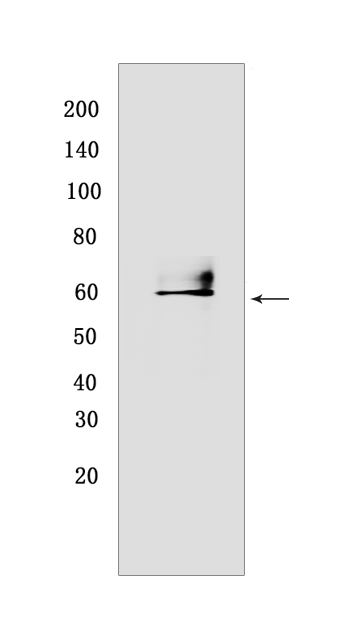
Western blot(SDS PAGE) analysis of extracts from HT-29 cells treated with a combination of the following treatments as indicated: Z-VAD (20 μM added 30 min prior to other compounds) Human Tumor Necrosis Factor-α(20 ng/ml 7 hr) SM-164 (100 nM 7 hr).Using P-RIP3 (S227) Rabbit mAb [75B5] at dilution of 1:1000 incubated at 4℃ over night.
Product information
Protein names :RIPK3,RIP3,RIPK3_HUMAN,Receptor-interacting serine/threonine-protein kinase 3
UniProtID :Q9Y572
MW(kDa) :46-62 kDa
Form :Liquid
Purification :Protein A purification
Host :Rabbit
Isotype :IgG
sensitivity :Endogenous
Reactivity :Human
- ApplicationDilution
- 免疫印迹(WB)1:1000-2000
- The optimal dilutions should be determined by the end user
Specificity :Antibody is produced by immunizing animals with a synthetic peptide at the sequence of Human Phospho-RIP3 (Ser227)
Storage :Antibody store in 10 mM PBS, 0.5mg/ml BSA, 50% glycerol. Shipped at 4°C. Store at-20°C or -80°C. Products are valid for one natural year of receipt.Avoid repeated freeze / thaw cycles.
WB Positive detected :HT-29 cells treated with a combination of the following treatments as indicated: Z-VAD (20 μM added 30 min prior to other compounds) Human Tumor Necrosis Factor-α(20 ng/ml 7 hr) SM-164 (100 nM 7 hr)
Function : Serine/threonine-protein kinase that activates necroptosis and apoptosis, two parallel forms of cell death (PubMed:19524512, PubMed:19524513, PubMed:22265413, PubMed:22265414, PubMed:22421439, PubMed:29883609, PubMed:32657447). Necroptosis, a programmed cell death process in response to death-inducing TNF-alpha family members, is triggered by RIPK3 following activation by ZBP1 (PubMed:19524512, PubMed:19524513, PubMed:22265413, PubMed:22265414, PubMed:22421439, PubMed:29883609, PubMed:32298652). Activated RIPK3 forms a necrosis-inducing complex and mediates phosphorylation of MLKL, promoting MLKL localization to the plasma membrane and execution of programmed necrosis characterized by calcium influx and plasma membrane damage (PubMed:19524512, PubMed:19524513, PubMed:22265413, PubMed:22265414, PubMed:22421439, PubMed:25316792, PubMed:29883609). In addition to TNF-induced necroptosis, necroptosis can also take place in the nucleus in response to orthomyxoviruses infection: following ZBP1 activation, which senses double-stranded Z-RNA structures, nuclear RIPK3 catalyzes phosphorylation and activation of MLKL, promoting disruption of the nuclear envelope and leakage of cellular DNA into the cytosol (By similarity). Also regulates apoptosis: apoptosis depends on RIPK1, FADD and CASP8, and is independent of MLKL and RIPK3 kinase activity (By similarity). Phosphorylates RIPK1: RIPK1 and RIPK3 undergo reciprocal auto- and trans-phosphorylation (PubMed:19524513). In some cell types, also able to restrict viral replication by promoting cell death-independent responses (By similarity). In response to Zika virus infection in neurons, promotes a cell death-independent pathway that restricts viral replication: together with ZBP1, promotes a death-independent transcriptional program that modifies the cellular metabolism via up-regulation expression of the enzyme ACOD1/IRG1 and production of the metabolite itaconate (By similarity). Itaconate inhibits the activity of succinate dehydrogenase, generating a metabolic state in neurons that suppresses replication of viral genomes (By similarity). RIPK3 binds to and enhances the activity of three metabolic enzymes: GLUL, GLUD1, and PYGL (PubMed:19498109). These metabolic enzymes may eventually stimulate the tricarboxylic acid cycle and oxidative phosphorylation, which could result in enhanced ROS production (PubMed:19498109).., (Microbial infection) In case of herpes simplex virus 1/HHV-1 infection, forms heteromeric amyloid structures with HHV-1 protein RIR1/ICP6 which may inhibit RIPK3-mediated necroptosis, thereby preventing host cell death pathway and allowing viral evasion..
Tissue specificity :Highly expressed in the pancreas. Detected at lower levels in heart, placenta, lung and kidney..,[Isoform 3]: Expression is significantly increased in colon and lung cancers..
Subcellular locationi :Cytoplasm, cytosol. Nucleus.
IMPORTANT: For western blots, incubate membrane with diluted primary antibody in 1% w/v BSA, 1X TBST at 4°C overnight.
-
About
-
Product
-
Law
-
Literature
-

WeChat scan code follow us