
PD-L1 Mouse mAb [C36S]Cat NO.: A90017
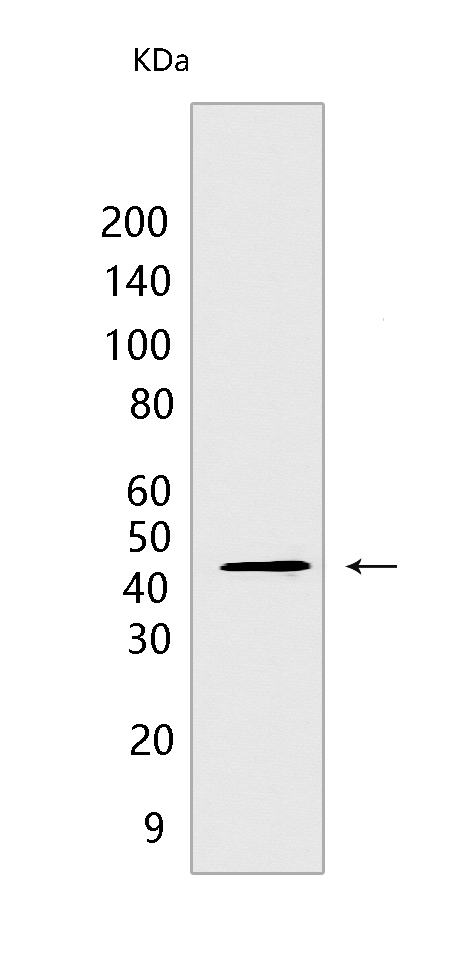
Western blot(SDS PAGE) analysis of extracts from KARPAS-299 cells.Using PD-L1 mouse mAb[C36S] at dilution of 1:1000 incubated at 4℃ over night.
Product information
Protein names :CD274,B7H1,PDCD1L1,PDCD1LG1,PDL1,PD1L1_HUMAN,Programmed cell death 1 ligand 1
UniProtID :Q9NZQ7
MASS(da) :33,275
MW(kDa) :40-50 kDa
Form :Liquid
Purification :Protein A purification
Host :mouse
Isotype :IgG
sensitivity :Endogenous
Reactivity :Human
- ApplicationDilution
- 免疫印迹(WB)1:1000-2000
- The optimal dilutions should be determined by the end user
Specificity :Antibody is produced by immunizing animals with a synthetic peptide at the sequence of Human PD-L1
Storage :Antibody store in 10 mM PBS, 0.5mg/ml BSA, 50% glycerol. Shipped at 4°C. Store at-20°C or -80°C. Products are valid for one natural year of receipt.Avoid repeated freeze / thaw cycles.
WB Positive detected :KARPAS-299 cells
Function : Plays a critical role in induction and maintenance of immune tolerance to self (PubMed:11015443, PubMed:28813417, PubMed:28813410). As a ligand for the inhibitory receptor PDCD1/PD-1, modulates the activation threshold of T-cells and limits T-cell effector response (PubMed:11015443, PubMed:28813417, PubMed:28813410). Through a yet unknown activating receptor, may costimulate T-cell subsets that predominantly produce interleukin-10 (IL10) (PubMed:10581077).., The PDCD1-mediated inhibitory pathway is exploited by tumors to attenuate anti-tumor immunity and escape destruction by the immune system, thereby facilitating tumor survival (PubMed:28813417, PubMed:28813410). The interaction with PDCD1/PD-1 inhibits cytotoxic T lymphocytes (CTLs) effector function (By similarity). The blockage of the PDCD1-mediated pathway results in the reversal of the exhausted T-cell phenotype and the normalization of the anti-tumor response, providing a rationale for cancer immunotherapy (By similarity)..
Tissue specificity :Highly expressed in the heart, skeletal muscle, placenta and lung. Weakly expressed in the thymus, spleen, kidney and liver. Expressed on activated T- and B-cells, dendritic cells, keratinocytes and monocytes..
Subcellular locationi :Cell membrane,Single-pass type I membrane protein. Early endosome membrane,Single-pass type I membrane protein. Recycling endosome membrane,Single-pass type I membrane protein.
IMPORTANT: For western blots, incubate membrane with diluted primary antibody in 1% w/v BSA, 1X TBST at 4°C overnight.
-
About
-
Product
-
Law
-
Literature
-

WeChat scan code follow us