
Cdc42 Rabbit pAbCat NO.: A58158
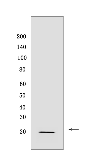
Western blot(SDS PAGE) analysis of extracts from 293T cells.Using Cdc42 Rabbit pAb at dilution of 1:1000 incubated at 4℃ over night.
Product information
Protein names :CDC42,CDC42_HUMAN,Cell division control protein 42 homolog
UniProtID :P60953
MASS(da) :21,259
MW(kDa) :21 kDa
Form :Liquid
Purification :peptide affinity chromatography
Host :Rabbit
Isotype :IgG
sensitivity :Endogenous
Reactivity :Human,Mouse,Rat
- ApplicationDilution
- 免疫印迹(WB)1:1000-2000
- The optimal dilutions should be determined by the end user
Specificity :Antibody is produced by immunizing animals with a synthetic peptide at the sequence of Human Cdc42
Storage :Antibody store in 10 mM PBS, 0.5mg/ml BSA, 50% glycerol. Shipped at 4°C. Store at-20°C or -80°C. Products are valid for one natural year of receipt.Avoid repeated freeze / thaw cycles.
WB Positive detected :293T cells
Function : Plasma membrane-associated small GTPase which cycles between an active GTP-bound and an inactive GDP-bound state. In active state binds to a variety of effector proteins to regulate cellular responses. Involved in epithelial cell polarization processes. Regulates the bipolar attachment of spindle microtubules to kinetochores before chromosome congression in metaphase (PubMed:15642749). Regulates cell migration (PubMed:17038317). In neurons, plays a role in the extension and maintenance of the formation of filopodia, thin and actin-rich surface projections (PubMed:14978216). Required for DOCK10-mediated spine formation in Purkinje cells and hippocampal neurons. In podocytes, facilitates filopodia and podosomes formation upon DOCK11-activation (PubMed:33523862). Upon activation by CaMKII, modulates dendritic spine structural plasticity by relaying CaMKII transient activation to synapse-specific, long-term signaling (By similarity). Also plays a role in phagocytosis through organization of the F-actin cytoskeleton associated with forming phagocytic cups (PubMed:26465210)..
Subcellular locationi :Cell membrane,Lipid-anchor,Cytoplasmic side. Cytoplasm, cytoskeleton, microtubule organizing center, centrosome. Cytoplasm, cytoskeleton, spindle. Midbody. Cell projection, dendrite.
IMPORTANT: For western blots, incubate membrane with diluted primary antibody in 1% w/v BSA, 1X TBST at 4°C overnight.
-
About
-
Product
-
Law
-
Literature
-

WeChat scan code follow us