
P-DDR1 (Y513) Rabbit mAb [8LY7]Cat NO.: A32493
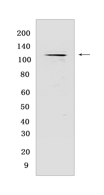
Western blot(SDS PAGE) analysis of extracts from T-47D cells treated with collagen (20 μg/ml 18h).Using P-DDR1 (Y513) Rabbit mAb [8LY7] at dilution of 1:1000 incubated at 4℃ over night.
Product information
Protein names :DDR1,CAK,EDDR1,NEP,NTRK4,PTK3A,RTK6,TRKE,DDR1_HUMAN,Epithelial discoidin domain-containing receptor 1
UniProtID :Q08345
MASS(da) :101,128
MW(kDa) :125 kDa
Form :Liquid
Purification :Protein A purification
Host :Rabbit
Isotype :IgG
sensitivity :Endogenous
Reactivity :Human
- ApplicationDilution
- 免疫印迹(WB)1:1000-2000
- The optimal dilutions should be determined by the end user
Specificity :Antibody is produced by immunizing animals with a synthetic peptide at the sequence of Human Phospho-DDR1 (Tyr513)
Storage :Antibody store in 10 mM PBS, 0.5mg/ml BSA, 50% glycerol. Shipped at 4°C. Store at-20°C or -80°C. Products are valid for one natural year of receipt.Avoid repeated freeze / thaw cycles.
WB Positive detected :T-47D cells treated with collagen (20 μg/ml 18h)
Function : Tyrosine kinase that functions as cell surface receptor for fibrillar collagen and regulates cell attachment to the extracellular matrix, remodeling of the extracellular matrix, cell migration, differentiation, survival and cell proliferation. Collagen binding triggers a signaling pathway that involves SRC and leads to the activation of MAP kinases. Regulates remodeling of the extracellular matrix by up-regulation of the matrix metalloproteinases MMP2, MMP7 and MMP9, and thereby facilitates cell migration and wound healing. Required for normal blastocyst implantation during pregnancy, for normal mammary gland differentiation and normal lactation. Required for normal ear morphology and normal hearing (By similarity). Promotes smooth muscle cell migration, and thereby contributes to arterial wound healing. Also plays a role in tumor cell invasion. Phosphorylates PTPN11..
Tissue specificity :Detected in T-47D, MDA-MB-175 and HBL-100 breast carcinoma cells, A-431 epidermoid carcinoma cells, SW48 and SNU-C2B colon carcinoma cells and Hs 294T melanoma cells (at protein level). Expressed at low levels in most adult tissues and is highest in the brain, lung, placenta and kidney. Lower levels of expression are detected in melanocytes, heart, liver, skeletal muscle and pancreas. Abundant in breast carcinoma cell lines. In the colonic mucosa, expressed in epithelia but not in the connective tissue of the lamina propria. In the thyroid gland, expressed in the epithelium of the thyroid follicles. In pancreas, expressed in the islets of Langerhans cells, but not in the surrounding epithelial cells of the exocrine pancreas. In kidney, expressed in the epithelia of the distal tubules. Not expressed in connective tissue, endothelial cells, adipose tissue, muscle cells or cells of hematopoietic origin..
Subcellular locationi :[Isoform 1]: Cell membrane,Single-pass type I membrane protein.,[Isoform 2]: Cell membrane,Single-pass type I membrane protein.,[Isoform 3]: Secreted.,[Isoform 4]: Cell membrane,Single-pass type I membrane protein.
IMPORTANT: For western blots, incubate membrane with diluted primary antibody in 1% w/v BSA, 1X TBST at 4°C overnight.
-
About
-
Product
-
Law
-
Literature
-

WeChat scan code follow us