
TIM3 Mouse mAb[BH72]Cat NO.: A68438
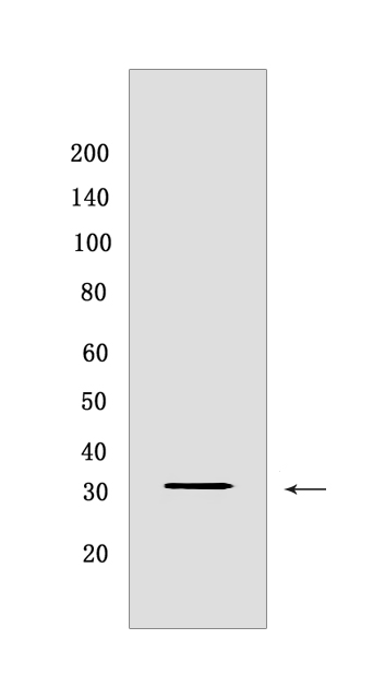
Western blot(SDS PAGE) analysis of extracts from Daudi cells lysates.Using TIM3 mouse mAb IgG [BH72] at dilution of 1:1000 incubated at 4℃ over night.
Product information
Protein names :HAVCR2,TIM3,TIMD3,HAVR2_HUMAN,Hepatitis A virus cellular receptor 2
UniProtID :Q8TDQ0
MASS(da) :33,394
MW(kDa) :33
Form :Liquid
Purification :Protein A purification
Host :mouse
Isotype :IgG
sensitivity :Endogenous
Reactivity :Human,Mouse,Rat
- ApplicationDilution
- 免疫印迹(WB)1:1000-2000
- The optimal dilutions should be determined by the end user
Specificity :Antibody is produced by immunizing animals with a synthetic peptide at the sequence of human TIM3.
Storage :Antibody store in 10 mM PBS, 0.5mg/ml BSA, 50% glycerol. Shipped at 4°C. Store at-20°C or -80°C. Products are valid for one natural year of receipt.Avoid repeated freeze / thaw cycles.
WB Positive detected :Daudi cells lysates
Function : Cell surface receptor implicated in modulating innate and adaptive immune responses. Generally accepted to have an inhibiting function. Reports on stimulating functions suggest that the activity may be influenced by the cellular context and/or the respective ligand (PubMed:24825777). Regulates macrophage activation (PubMed:11823861). Inhibits T-helper type 1 lymphocyte (Th1)-mediated auto- and alloimmune responses and promotes immunological tolerance (PubMed:14556005). In CD8+ cells attenuates TCR-induced signaling, specifically by blocking NF-kappaB and NFAT promoter activities resulting in the loss of IL-2 secretion. The function may implicate its association with LCK proposed to impair phosphorylation of TCR subunits, and/or LGALS9-dependent recruitment of PTPRC to the immunological synapse (PubMed:24337741, PubMed:26492563). In contrast, shown to activate TCR-induced signaling in T-cells probably implicating ZAP70, LCP2, LCK and FYN (By similarity). Expressed on Treg cells can inhibit Th17 cell responses (PubMed:24838857). Receptor for LGALS9 (PubMed:16286920, PubMed:24337741). Binding to LGALS9 is believed to result in suppression of T-cell responses,the resulting apoptosis of antigen-specific cells may implicate HAVCR2 phosphorylation and disruption of its association with BAG6. Binding to LGALS9 is proposed to be involved in innate immune response to intracellular pathogens. Expressed on Th1 cells interacts with LGALS9 expressed on Mycobacterium tuberculosis-infected macrophages to stimulate antibactericidal activity including IL-1 beta secretion and to restrict intracellular bacterial growth (By similarity). However, the function as receptor for LGALS9 has been challenged (PubMed:23555261). Also reported to enhance CD8+ T-cell responses to an acute infection such as by Listeria monocytogenes (By similarity). Receptor for phosphatidylserine (PtSer),PtSer-binding is calcium-dependent. May recognize PtSer on apoptotic cells leading to their phagocytosis. Mediates the engulfment of apoptotic cells by dendritic cells. Expressed on T-cells, promotes conjugation but not engulfment of apoptotic cells. Expressed on dendritic cells (DCs) positively regulates innate immune response and in synergy with Toll-like receptors promotes secretion of TNF-alpha. In tumor-imfiltrating DCs suppresses nucleic acid-mediated innate immune repsonse by interaction with HMGB1 and interfering with nucleic acid-sensing and trafficking of nucleid acids to endosomes (By similarity). Expressed on natural killer (NK) cells acts as a coreceptor to enhance IFN-gamma production in response to LGALS9 (PubMed:22323453). In contrast, shown to suppress NK cell-mediated cytotoxicity (PubMed:22383801). Negatively regulates NK cell function in LPS-induced endotoxic shock (By similarity)..
Tissue specificity :Expressed in T-helper type 1 (Th1) lymphocytes. Expressed on regulatory T (Treg) cells after TCR stimulation. Expressed in dendritic cells and natural killer (NK) cells. Expressed in epithelial tissues. Expression is increased on CD4+ and CD8+ T-cells in chronic hepatitis C virus (HCV) infection. In progressive HIV-1 infection, expression is up-regulated on HIV-1-specific CD8 T-cells..
Subcellular locationi :Membrane,Single-pass type I membrane protein. Cell junction. Cell membrane.
IMPORTANT: For western blots, incubate membrane with diluted primary antibody in 1% w/v BSA, 1X TBST at 4°C overnight.
-
About
-
Product
-
Law
-
Literature
-

WeChat scan code follow us