
B7-H3 Mouse mAb[4G53]Cat NO.: A56143
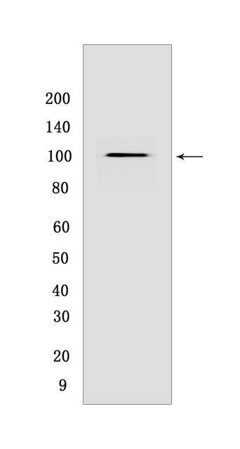
Western blot(SDS PAGE) analysis of extracts from HEK-293 cells lysates.Using B7-H3 mouse mAb IgG [4G53] at dilution of 1:1000 incubated at 4℃ over night.
Product information
Protein names :CD276,B7H3,PSEC0249,UNQ309/PRO352,CD276_HUMAN,CD276 antigen
UniProtID :Q5ZPR3
MASS(da) :57,235
MW(kDa) :100
Form :Liquid
Purification :Protein A purification
Host :mouse
Isotype :IgG
sensitivity :Endogenous
Reactivity :Human,Mouse
- ApplicationDilution
- 免疫印迹(WB)1:1000-2000,
- 免疫组化(IHC)1:100
- 免疫荧光(ICC/IF) 1:100
- The optimal dilutions should be determined by the end user
Specificity :Antibody is produced by immunizing animals with a synthetic peptide at the sequence of human B7-H3.
Storage :Antibody store in 10 mM PBS, 0.5mg/ml BSA, 50% glycerol. Shipped at 4°C. Store at-20°C or -80°C. Products are valid for one natural year of receipt.Avoid repeated freeze / thaw cycles.
WB Positive detected :HEK-293 cells lysates
Function : May participate in the regulation of T-cell-mediated immune response. May play a protective role in tumor cells by inhibiting natural-killer mediated cell lysis as well as a role of marker for detection of neuroblastoma cells. May be involved in the development of acute and chronic transplant rejection and in the regulation of lymphocytic activity at mucosal surfaces. Could also play a key role in providing the placenta and fetus with a suitable immunological environment throughout pregnancy. Both isoform 1 and isoform 2 appear to be redundant in their ability to modulate CD4 T-cell responses. Isoform 2 is shown to enhance the induction of cytotoxic T-cells and selectively stimulates interferon gamma production in the presence of T-cell receptor signaling..
Tissue specificity :Ubiquitous but not detectable in peripheral blood lymphocytes or granulocytes. Weakly expressed in resting monocytes. Expressed in dendritic cells derived from monocytes. Expressed in epithelial cells of sinonasal tissue. Expressed in extravillous trophoblast cells and Hofbauer cells of the first trimester placenta and term placenta..
Subcellular locationi :Membrane,Single-pass type I membrane protein.
IMPORTANT: For western blots, incubate membrane with diluted primary antibody in 1% w/v BSA, 1X TBST at 4°C overnight.
-
About
-
Product
-
Law
-
Literature
-

WeChat scan code follow us