
Phospho-FLT3 (Tyr591) Rabbit mAb[AZM5]Cat NO.: A45239
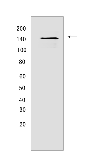
Western blot(SDS PAGE) analysis of extracts from SEM cell .Using Phospho-FLT3 (Tyr591) Rabbit mAb IgG [AZM5] at dilution of 1:1000 incubated at 4℃ over night.
Product information
Protein names :FLT3,CD135,FLK2,STK1,FLT3_HUMAN,Receptor-type tyrosine-protein kinase FLT3
UniProtID :P36888
MASS(da) :112,903
MW(kDa) : 160
Form :Liquid
Purification :Protein A purification
Host :Rabbit
Isotype :IgG
sensitivity :Endogenous
Reactivity :Human,Mouse
- ApplicationDilution
- 免疫印迹(WB)1:1000-2000
- The optimal dilutions should be determined by the end user
Specificity :Antibody is produced by immunizing animals with a synthetic peptide corresponding to residues surrounding Tyr591 of human FLT3
Storage :Antibody store in 10 mM PBS, 0.5mg/ml BSA, 50% glycerol. Shipped at 4°C. Store at-20°C or -80°C. Products are valid for one natural year of receipt.Avoid repeated freeze / thaw cycles.
WB Positive detected :SEM cell
Function : Tyrosine-protein kinase that acts as cell-surface receptor for the cytokine FLT3LG and regulates differentiation, proliferation and survival of hematopoietic progenitor cells and of dendritic cells. Promotes phosphorylation of SHC1 and AKT1, and activation of the downstream effector MTOR. Promotes activation of RAS signaling and phosphorylation of downstream kinases, including MAPK1/ERK2 and/or MAPK3/ERK1. Promotes phosphorylation of FES, FER, PTPN6/SHP, PTPN11/SHP-2, PLCG1, and STAT5A and/or STAT5B. Activation of wild-type FLT3 causes only marginal activation of STAT5A or STAT5B. Mutations that cause constitutive kinase activity promote cell proliferation and resistance to apoptosis via the activation of multiple signaling pathways..
Tissue specificity :Detected in bone marrow, in hematopoietic stem cells, in myeloid progenitor cells and in granulocyte/macrophage progenitor cells (at protein level). Detected in bone marrow, liver, thymus, spleen and lymph node, and at low levels in kidney and pancreas. Highly expressed in T-cell leukemia..
Subcellular locationi :Membrane,Single-pass type I membrane protein. Endoplasmic reticulum lumen.
IMPORTANT: For western blots, incubate membrane with diluted primary antibody in 1% w/v BSA, 1X TBST at 4°C overnight.
-
About
-
Product
-
Law
-
Literature
-

WeChat scan code follow us