
Fgr Rabbit pAbCat NO.: A96004
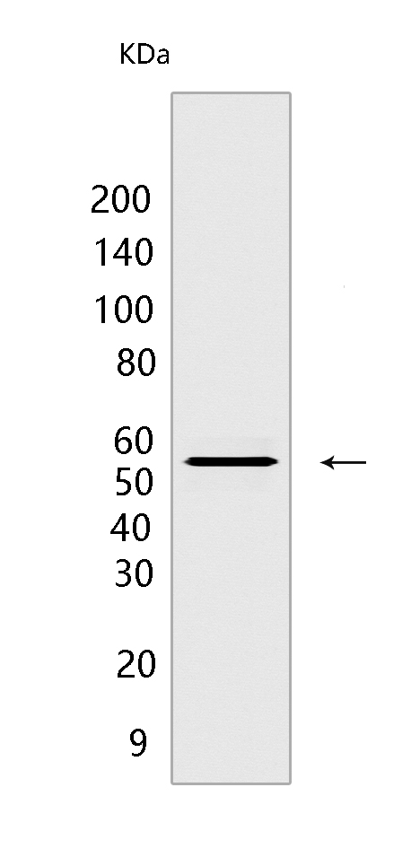
Western blot(SDS PAGE) analysis of extracts from Raji cells.Using Fgr Rabbit pAb at dilution of 1:1000 incubated at 4℃ over night.
Product information
Protein names :FGR,SRC2,FGR_HUMAN,Tyrosine-protein kinase Fgr oncogene homolog)
UniProtID :P09769
MASS(da) :59,479
MW(kDa) :56 kDa
Form :Liquid
Purification :peptide affinity chromatography
Host :Rabbit
Isotype :IgG
sensitivity :Endogenous
Reactivity :Human
- ApplicationDilution
- 免疫印迹(WB)1:1000-2000
- The optimal dilutions should be determined by the end user
Specificity :Antibody is produced by immunizing animals with a synthetic peptide at the sequence of Human Fgr
Storage :Antibody store in 10 mM PBS, 0.5mg/ml BSA, 50% glycerol. Shipped at 4°C. Store at-20°C or -80°C. Products are valid for one natural year of receipt.Avoid repeated freeze / thaw cycles.
WB Positive detected :Raji cells
Function : Non-receptor tyrosine-protein kinase that transmits signals from cell surface receptors devoid of kinase activity and contributes to the regulation of immune responses, including neutrophil, monocyte, macrophage and mast cell functions, cytoskeleton remodeling in response to extracellular stimuli, phagocytosis, cell adhesion and migration. Promotes mast cell degranulation, release of inflammatory cytokines and IgE-mediated anaphylaxis. Acts downstream of receptors that bind the Fc region of immunoglobulins, such as MS4A2/FCER1B, FCGR2A and/or FCGR2B. Acts downstream of ITGB1 and ITGB2, and regulates actin cytoskeleton reorganization, cell spreading and adhesion. Depending on the context, activates or inhibits cellular responses. Functions as negative regulator of ITGB2 signaling, phagocytosis and SYK activity in monocytes. Required for normal ITGB1 and ITGB2 signaling, normal cell spreading and adhesion in neutrophils and macrophages. Functions as positive regulator of cell migration and regulates cytoskeleton reorganization via RAC1 activation. Phosphorylates SYK (in vitro) and promotes SYK-dependent activation of AKT1 and MAP kinase signaling. Phosphorylates PLD2 in antigen-stimulated mast cells, leading to PLD2 activation and the production of the signaling molecules lysophosphatidic acid and diacylglycerol. Promotes activation of PIK3R1. Phosphorylates FASLG, and thereby regulates its ubiquitination and subsequent internalization. Phosphorylates ABL1. Promotes phosphorylation of CBL, CTTN, PIK3R1, PTK2/FAK1, PTK2B/PYK2 and VAV2. Phosphorylates HCLS1 that has already been phosphorylated by SYK, but not unphosphorylated HCLS1. Together with CLNK, it acts as a negative regulator of natural killer cell-activating receptors and inhibits interferon-gamma production (By similarity)..
Tissue specificity :Detected in neutrophils, monocytes and natural killer cells (at protein level). Detected in monocytes and large lymphocytes..
Subcellular locationi :Cell membrane,Lipid-anchor,Cytoplasmic side. Cell membrane,Peripheral membrane protein,Cytoplasmic side. Cell projection, ruffle membrane. Cytoplasm, cytosol. Cytoplasm, cytoskeleton. Mitochondrion inner membrane. Mitochondrion intermembrane space.
IMPORTANT: For western blots, incubate membrane with diluted primary antibody in 1% w/v BSA, 1X TBST at 4°C overnight.
-
About
-
Product
-
Law
-
Literature
-

WeChat scan code follow us