
Akt3 Rabbit mAb [69IU]Cat NO.: A45235
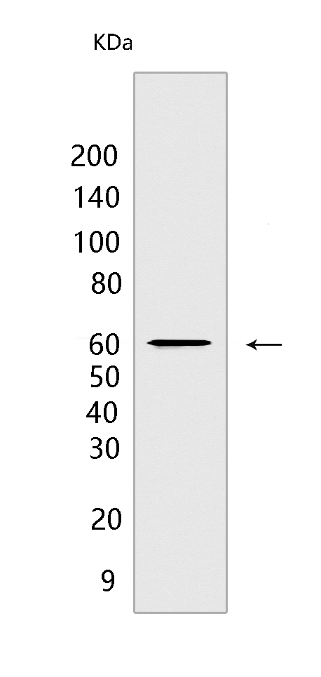
Western blot(SDS PAGE) analysis of extracts from A172 cells.Using Akt3 Rabbit mAb [69IU] at dilution of 1:1000 incubated at 4℃ over night.
Product information
Protein names :AKT3,PKBG,AKT3_HUMAN,RAC-gamma serine/threonine-protein kinase
UniProtID :Q9Y243
MASS(da) :55,775
MW(kDa) :60 kDa
Form :Liquid
Purification :Protein A purification
Host :Rabbit
Isotype :IgG
sensitivity :Endogenous
Reactivity :Human,Mouse,Rat
- ApplicationDilution
- 免疫印迹(WB)1:1000-2000
- The optimal dilutions should be determined by the end user
Specificity :Antibody is produced by immunizing animals with a synthetic peptide at the sequence of Human Akt3
Storage :Antibody store in 10 mM PBS, 0.5mg/ml BSA, 50% glycerol. Shipped at 4°C. Store at-20°C or -80°C. Products are valid for one natural year of receipt.Avoid repeated freeze / thaw cycles.
WB Positive detected :A172 cells
Function : AKT3 is one of 3 closely related serine/threonine-protein kinases (AKT1, AKT2 and AKT3) called the AKT kinase, and which regulate many processes including metabolism, proliferation, cell survival, growth and angiogenesis. This is mediated through serine and/or threonine phosphorylation of a range of downstream substrates. Over 100 substrate candidates have been reported so far, but for most of them, no isoform specificity has been reported. AKT3 is the least studied AKT isoform. It plays an important role in brain development and is crucial for the viability of malignant glioma cells. AKT3 isoform may also be the key molecule in up-regulation and down-regulation of MMP13 via IL13. Required for the coordination of mitochondrial biogenesis with growth factor-induced increases in cellular energy demands. Down-regulation by RNA interference reduces the expression of the phosphorylated form of BAD, resulting in the induction of caspase-dependent apoptosis..
Tissue specificity :In adult tissues, it is highly expressed in brain, lung and kidney, but weakly in heart, testis and liver. In fetal tissues, it is highly expressed in heart, liver and brain and not at all in kidney.
Subcellular locationi :Nucleus. Cytoplasm. Membrane,Peripheral membrane protein.
IMPORTANT: For western blots, incubate membrane with diluted primary antibody in 1% w/v BSA, 1X TBST at 4°C overnight.
-
About
-
Product
-
Law
-
Literature
-

WeChat scan code follow us