
ATP5A1 Mouse mAb[4FO7]Cat NO.: A58643
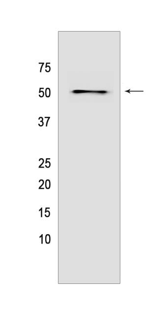
Western blot(SDS PAGE) analysis of extracts from MCF-7 cells.Using ATP5A1 Mouse mAb IgG [4FO7] at dilution of 1:1000 incubated at 4℃ over night.
Product information
Protein names :ATP5F1A,ATP5A,ATP5A1,ATP5AL2,ATPM,ATPA_HUMAN,ATP synthase subunit alpha, mitochondrial
UniProtID :P25705
MASS(da) :59,751
MW(kDa) :50kDa
Form :Liquid
Purification :Protein A purification
Host :Mouse
Isotype :IgG
sensitivity :Endogenous
Reactivity :Human,Mouse,Rat
- ApplicationDilution
- 免疫印迹(WB)1:1000-2000
- 免疫组化(IHC)1:100
- 免疫荧光(ICC/IF) 1:100,
- The optimal dilutions should be determined by the end user
Specificity :Antibody is produced by immunizing animals with a synthetic peptide of human ATP5A1.
Storage :Antibody store in 10 mM PBS, 0.5mg/ml BSA, 50% glycerol. Shipped at 4°C. Store at-20°C or -80°C. Products are valid for one natural year of receipt.Avoid repeated freeze / thaw cycles.
WB Positive detected :MCF-7 cells
Function : Mitochondrial membrane ATP synthase (F(1)F(0) ATP synthase or Complex V) produces ATP from ADP in the presence of a proton gradient across the membrane which is generated by electron transport complexes of the respiratory chain. F-type ATPases consist of two structural domains, F(1) - containing the extramembraneous catalytic core, and F(0) - containing the membrane proton channel, linked together by a central stalk and a peripheral stalk. During catalysis, ATP synthesis in the catalytic domain of F(1) is coupled via a rotary mechanism of the central stalk subunits to proton translocation. Subunits alpha and beta form the catalytic core in F(1). Rotation of the central stalk against the surrounding alpha(3)beta(3) subunits leads to hydrolysis of ATP in three separate catalytic sites on the beta subunits. Subunit alpha does not bear the catalytic high-affinity ATP-binding sites (By similarity). Binds the bacterial siderophore enterobactin and can promote mitochondrial accumulation of enterobactin-derived iron ions (PubMed:30146159)..
Tissue specificity :Fetal lung, heart, liver, gut and kidney. Expressed at higher levels in the fetal brain, retina and spinal cord..
Subcellular locationi :Mitochondrion. Mitochondrion inner membrane,Peripheral membrane protein,Matrix side. Cell membrane,Peripheral membrane protein,Extracellular side.
IMPORTANT: For western blots, incubate membrane with diluted primary antibody in 1% w/v BSA, 1X TBST at 4°C overnight.
-
About
-
Product
-
Law
-
Literature
-

WeChat scan code follow us