
ATG5 Mouse mAb[G8H4]Cat NO.: A82026
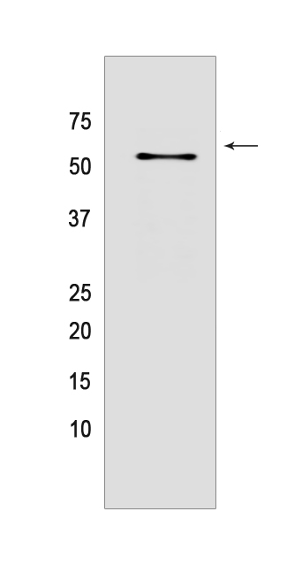
Western blot(SDS PAGE) analysis of extracts from HEK-293 cells.Using ATG5 Mouse mAb IgG [G8H4] at dilution of 1:1000 incubated at 4℃ over night.
Product information
Protein names :ATG5,APG5L,ASP,ATG5_HUMAN,Autophagy protein 5
UniProtID :Q9H1Y0
MASS(da) :32,447
MW(kDa) :50-55and40-45kDa
Form :Liquid
Purification :Protein A purification
Host :Mouse
Isotype :IgG
sensitivity :Endogenous
Reactivity :Human,Mouse,Rat
- ApplicationDilution
- 免疫印迹(WB)1:1000-2000
- 免疫组化(IHC)1:100
- The optimal dilutions should be determined by the end user
Specificity :Antibody is produced by immunizing animals with a synthetic peptide of human ATG5.
Storage :Antibody store in 10 mM PBS, 0.5mg/ml BSA, 50% glycerol. Shipped at 4°C. Store at-20°C or -80°C. Products are valid for one natural year of receipt.Avoid repeated freeze / thaw cycles.
WB Positive detected :HEK-293 cells
Function : Involved in autophagic vesicle formation. Conjugation with ATG12, through a ubiquitin-like conjugating system involving ATG7 as an E1-like activating enzyme and ATG10 as an E2-like conjugating enzyme, is essential for its function. The ATG12-ATG5 conjugate acts as an E3-like enzyme which is required for lipidation of ATG8 family proteins and their association to the vesicle membranes. Involved in mitochondrial quality control after oxidative damage, and in subsequent cellular longevity. Plays a critical role in multiple aspects of lymphocyte development and is essential for both B and T lymphocyte survival and proliferation. Required for optimal processing and presentation of antigens for MHC II. Involved in the maintenance of axon morphology and membrane structures, as well as in normal adipocyte differentiation. Promotes primary ciliogenesis through removal of OFD1 from centriolar satellites and degradation of IFT20 via the autophagic pathway.., May play an important role in the apoptotic process, possibly within the modified cytoskeleton. Its expression is a relatively late event in the apoptotic process, occurring downstream of caspase activity. Plays a crucial role in IFN-gamma-induced autophagic cell death by interacting with FADD.., (Microbial infection) May act as a proviral factor. In association with ATG12, negatively regulates the innate antiviral immune response by impairing the type I IFN production pathway upon vesicular stomatitis virus (VSV) infection (PubMed:17709747). Required for the translation of incoming hepatitis C virus (HCV) RNA and, thereby, for initiation of HCV replication, but not required once infection is established (PubMed:19666601)..
Tissue specificity :Ubiquitous. The mRNA is present at similar levels in viable and apoptotic cells, whereas the protein is dramatically highly expressed in apoptotic cells.
Subcellular locationi :Cytoplasm. Preautophagosomal structure membrane,Peripheral membrane protein.
IMPORTANT: For western blots, incubate membrane with diluted primary antibody in 1% w/v BSA, 1X TBST at 4°C overnight.
-
About
-
Product
-
Law
-
Literature
-

WeChat scan code follow us