
Myocilin Mouse mAb[436Z]Cat NO.: A91795
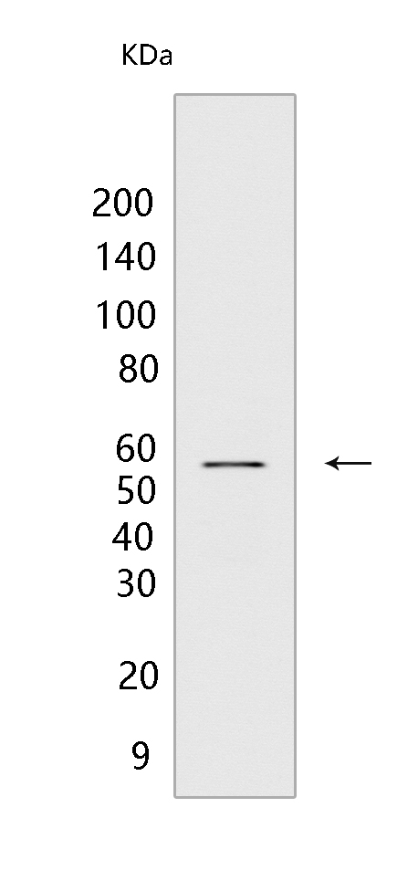
Western blot(SDS PAGE) analysis of extracts from human skeletal muscle tissue.Using Myocilin Mouse mAb IgG [436Z] at dilution of 1:1000 incubated at 4℃ over night.
Product information
Protein names :MYOC,GLC1A,TIGR,MYOC_HUMAN,Myocilin [Cleaved into: Myocilin, N-terminal fragment ,Myocilin, C-terminal fragment ]
UniProtID :Q99972
MASS(da) :56,972
MW(kDa) :57kda
Form :Liquid
Purification :Protein A purification
Host :Mouse
Isotype :IgG
sensitivity :Endogenous
Reactivity :Human,Mouse,Rat
- ApplicationDilution
- 免疫印迹(WB)1:1000-2000
- 免疫组化(IHC)1:100
- The optimal dilutions should be determined by the end user
Specificity :Antibody is produced by immunizing animals with a synthetic peptide of human Myocilin.
Storage :Antibody store in 10 mM PBS, 0.5mg/ml BSA, 50% glycerol. Shipped at 4°C. Store at-20°C or -80°C. Products are valid for one natural year of receipt.Avoid repeated freeze / thaw cycles.
WB Positive detected :human skeletal muscle tissue
Function : Secreted glycoprotein regulating the activation of different signaling pathways in adjacent cells to control different processes including cell adhesion, cell-matrix adhesion, cytoskeleton organization and cell migration. Promotes substrate adhesion, spreading and formation of focal contacts. Negatively regulates cell-matrix adhesion and stress fiber assembly through Rho protein signal transduction. Modulates the organization of actin cytoskeleton by stimulating the formation of stress fibers through interactions with components of Wnt signaling pathways. Promotes cell migration through activation of PTK2 and the downstream phosphatidylinositol 3-kinase signaling. Plays a role in bone formation and promotes osteoblast differentiation in a dose-dependent manner through mitogen-activated protein kinase signaling. Mediates myelination in the peripheral nervous system through ERBB2/ERBB3 signaling. Plays a role as a regulator of muscle hypertrophy through the components of dystrophin-associated protein complex. Involved in positive regulation of mitochondrial depolarization. Plays a role in neurite outgrowth. May participate in the obstruction of fluid outflow in the trabecular meshwork..
Tissue specificity :Detected in aqueous humor (PubMed:12697062). Detected in the eye (at protein level) (PubMed:11431441). Widely expressed. Highly expressed in various types of muscle, ciliary body, papillary sphincter, skeletal muscle, heart, and bone marrow-derived mesenchymal stem cells. Expressed predominantly in the retina. In normal eyes, found in the inner uveal meshwork region and the anterior portion of the meshwork. In contrast, in many glaucomatous eyes, it is found in more regions of the meshwork and seems to be expressed at higher levels than in normal eyes, regardless of the type or clinical severity of glaucoma. The myocilin 35 kDa fragment is detected in aqueous humor and to a lesser extent in iris and ciliary body..
Subcellular locationi :Secreted. Golgi apparatus. Cytoplasmic vesicle. Secreted, extracellular space. Secreted, extracellular space, extracellular matrix. Secreted, extracellular exosome. Mitochondrion. Mitochondrion intermembrane space. Mitochondrion inner membrane. Mitochondrion outer membrane. Rough endoplasmic reticulum. Cell projection. Cell projection, cilium.
IMPORTANT: For western blots, incubate membrane with diluted primary antibody in 1% w/v BSA, 1X TBST at 4°C overnight.
-
About
-
Product
-
Law
-
Literature
-

WeChat scan code follow us