
SET Mouse mAb[5720]Cat NO.: A95753
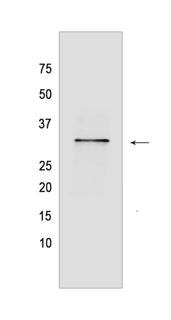
Western blot(SDS PAGE) analysis of extracts from Jurkat cells.Using SET Mouse mAb IgG [5720] at dilution of 1:1000 incubated at 4℃ over night.
Product information
Protein names :SET,SET_HUMAN,Protein SET
UniProtID :Q01105
MASS(da) :33,489
MW(kDa) :33kda
Form :Liquid
Purification :Protein A purification
Host :Mouse
Isotype :IgG
sensitivity :Endogenous
Reactivity :Human,Mouse,Rat
- ApplicationDilution
- 免疫印迹(WB)1:1000-2000
- The optimal dilutions should be determined by the end user
Specificity :Antibody is produced by immunizing animals with a synthetic peptide of human SET.
Storage :Antibody store in 10 mM PBS, 0.5mg/ml BSA, 50% glycerol. Shipped at 4°C. Store at-20°C or -80°C. Products are valid for one natural year of receipt.Avoid repeated freeze / thaw cycles.
WB Positive detected :Jurkat cells
Function : Multitasking protein, involved in apoptosis, transcription, nucleosome assembly and histone chaperoning. Isoform 2 anti-apoptotic activity is mediated by inhibition of the GZMA-activated DNase, NME1. In the course of cytotoxic T-lymphocyte (CTL)-induced apoptosis, GZMA cleaves SET, disrupting its binding to NME1 and releasing NME1 inhibition. Isoform 1 and isoform 2 are potent inhibitors of protein phosphatase 2A. Isoform 1 and isoform 2 inhibit EP300/CREBBP and PCAF-mediated acetylation of histones (HAT) and nucleosomes, most probably by masking the accessibility of lysines of histones to the acetylases. The predominant target for inhibition is histone H4. HAT inhibition leads to silencing of HAT-dependent transcription and prevents active demethylation of DNA. Both isoforms stimulate DNA replication of the adenovirus genome complexed with viral core proteins,however, isoform 2 specific activity is higher..
Tissue specificity :Widely expressed. Low levels in quiescent cells during serum starvation, contact inhibition or differentiation. Highly expressed in Wilms' tumor.
Subcellular locationi :Cytoplasm, cytosol. Endoplasmic reticulum. Nucleus, nucleoplasm.
IMPORTANT: For western blots, incubate membrane with diluted primary antibody in 1% w/v BSA, 1X TBST at 4°C overnight.
-
About
-
Product
-
Law
-
Literature
-

WeChat scan code follow us