
SOCS3 Mouse mAb[IJC2]Cat NO.: A72191
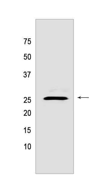
Western blot(SDS PAGE) analysis of extracts from Daudi cells.Using SOCS3 Mouse mAb IgG [IJC2] at dilution of 1:1000 incubated at 4℃ over night.
Product information
Protein names :SOCS3,CIS3,SSI3,SOCS3_HUMAN,Suppressor of cytokine signaling 3
UniProtID :O14543
MASS(da) :24,770
MW(kDa) :25kDa
Form :Liquid
Purification :Protein A purification
Host :Mouse
Isotype :IgG
sensitivity :Endogenous
Reactivity :Human,Mouse
- ApplicationDilution
- 免疫印迹(WB)1:1000-2000
- 免疫组化(IHC)1:100
- 免疫荧光(ICC/IF) 1:100,
- The optimal dilutions should be determined by the end user
Specificity :Antibody is produced by immunizing animals with a synthetic peptide of human SOCS3.
Storage :Antibody store in 10 mM PBS, 0.5mg/ml BSA, 50% glycerol. Shipped at 4°C. Store at-20°C or -80°C. Products are valid for one natural year of receipt.Avoid repeated freeze / thaw cycles.
WB Positive detected :Daudi cells
Function : SOCS family proteins form part of a classical negative feedback system that regulates cytokine signal transduction. SOCS3 is involved in negative regulation of cytokines that signal through the JAK/STAT pathway. Inhibits cytokine signal transduction by binding to tyrosine kinase receptors including IL6ST/gp130, LIF, erythropoietin, insulin, IL12, GCSF and leptin receptors. Binding to JAK2 inhibits its kinase activity and regulates IL6 signaling. Suppresses fetal liver erythropoiesis. Regulates onset and maintenance of allergic responses mediated by T-helper type 2 cells (By similarity). Probable substrate recognition component of a SCF-like ECS (Elongin BC-CUL2/5-SOCS-box protein) E3 ubiquitin-protein ligase complex which mediates the ubiquitination and subsequent proteasomal degradation of target proteins (PubMed:15601820)..
Tissue specificity :Widely expressed with high expression in heart, placenta, skeletal muscle, peripheral blood leukocytes, fetal and adult lung, and fetal liver and kidney. Lower levels in thymus.
IMPORTANT: For western blots, incubate membrane with diluted primary antibody in 1% w/v BSA, 1X TBST at 4°C overnight.
-
About
-
Product
-
Law
-
Literature
-

WeChat scan code follow us