
TLR4 Mouse mAb[F6MH]Cat NO.: A81340
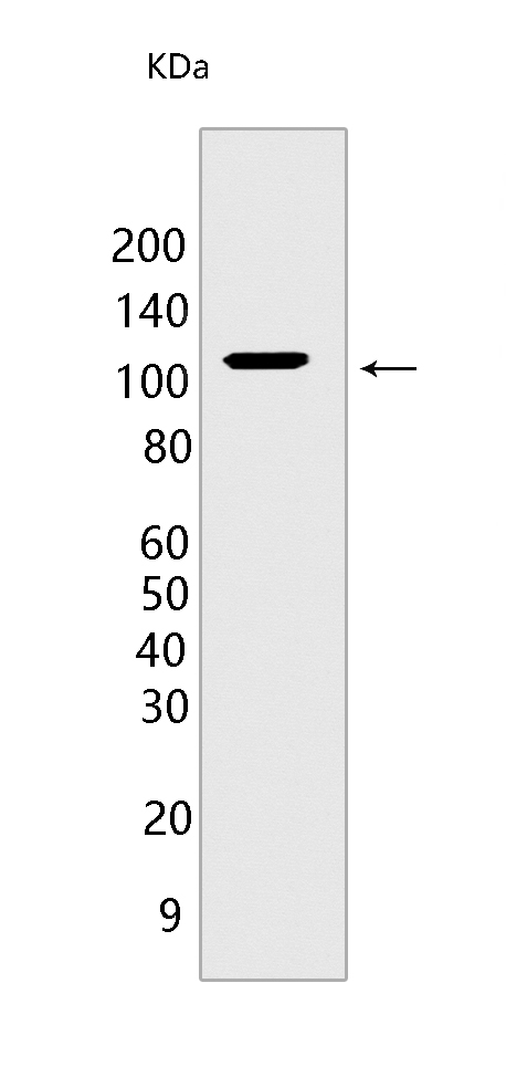
Western blot(SDS PAGE) analysis of extracts from RAW 264.7 cells.Using TLR4 Mouse mAb IgG [F6MH] at dilution of 1:1000 incubated at 4℃ over night.
Product information
Protein names :TLR4,TLR4_HUMAN,Toll-like receptor 4
UniProtID :O00206
MASS(da) :95,680
MW(kDa) :110 kda
Form :Liquid
Purification :Protein A purification
Host :Mouse
Isotype :IgG
sensitivity :Endogenous
Reactivity :Human,Mouse,Rat
- ApplicationDilution
- 免疫印迹(WB)1:1000-2000
- The optimal dilutions should be determined by the end user
Specificity :Antibody is produced by immunizing animals with a synthetic peptide of human TLR4
Storage :Antibody store in 10 mM PBS, 0.5mg/ml BSA, 50% glycerol. Shipped at 4°C. Store at-20°C or -80°C. Products are valid for one natural year of receipt.Avoid repeated freeze / thaw cycles.
WB Positive detected :RAW 264.7 cells
Function : Cooperates with LY96 and CD14 to mediate the innate immune response to bacterial lipopolysaccharide (LPS) (PubMed:27022195). Acts via MYD88, TIRAP and TRAF6, leading to NF-kappa-B activation, cytokine secretion and the inflammatory response (PubMed:9237759, PubMed:10835634, PubMed:27022195,PubMed:21393102). Also involved in LPS-independent inflammatory responses triggered by free fatty acids, such as palmitate, and Ni(2+). Responses triggered by Ni(2+) require non-conserved histidines and are, therefore, species-specific (PubMed:20711192). Both M.tuberculosis HSP70 (dnaK) and HSP65 (groEL-2) act via this protein to stimulate NF-kappa-B expression (PubMed:15809303). In complex with TLR6, promotes sterile inflammation in monocytes/macrophages in response to oxidized low-density lipoprotein (oxLDL) or amyloid-beta 42. In this context, the initial signal is provided by oxLDL- or amyloid-beta 42-binding to CD36. This event induces the formation of a heterodimer of TLR4 and TLR6, which is rapidly internalized and triggers inflammatory response, leading to the NF-kappa-B-dependent production of CXCL1, CXCL2 and CCL9 cytokines, via MYD88 signaling pathway, and CCL5 cytokine, via TICAM1 signaling pathway, as well as IL1B secretion. Binds electronegative LDL (LDL(-)) and mediates the cytokine release induced by LDL(-) (PubMed:23880187). Stimulation of monocytes in vitro with M.tuberculosis PstS1 induces p38 MAPK and ERK1/2 activation primarily via TLR2, but also partially via this receptor (PubMed:16622205, PubMed:10835634, PubMed:15809303, PubMed:17478729, PubMed:20037584, PubMed:20711192, PubMed:23880187, PubMed:27022195, PubMed:9237759). Activated by the signaling pathway regulator NMI which acts as damage-associated molecular patterns (DAMPs) in response to cell injury or pathogen invasion, therefore promoting nuclear factor NF-kappa-B activation (PubMed:29038465)..
Tissue specificity :Highly expressed in placenta, spleen and peripheral blood leukocytes (PubMed:9435236, PubMed:9237759). Detected in monocytes, macrophages, dendritic cells and several types of T-cells (PubMed:9237759, PubMed:27022195)..
Subcellular locationi :Cell membrane,Single-pass type I membrane protein. Early endosome. Cell projection, ruffle.
IMPORTANT: For western blots, incubate membrane with diluted primary antibody in 1% w/v BSA, 1X TBST at 4°C overnight.
-
About
-
Product
-
Law
-
Literature
-

WeChat scan code follow us