
VE-cadherin Mouse mAb [A9T2]Cat NO.: A44337
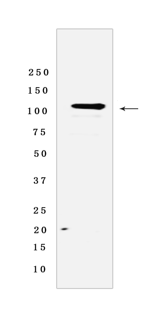
Western blot(SDS PAGE) analysis of extracts from human placenta tissue lysates. Using VE-cadherin Mouse mAb [A9T2] at dilution of 1:1000 incubated at 4℃ over night.
Product information
Protein names :CDH5,CADH5_HUMAN,Cadherin-5
UniProtID :P33151
MASS(da) :87,528
MW(kDa) :125 kDa
Form :Liquid
Purification :Protein A purification
Host :Mouse IgG
Isotype :IgG
sensitivity :Endogenous
Reactivity :Human
- ApplicationDilution
- 免疫印迹(WB)1:1000-2000
- 免疫组化(IHC)1:100
- The optimal dilutions should be determined by the end user
Specificity :Antibody is produced by immunizing animals with a synthetic peptide of Human VE-cadherin.
Storage :Antibody store in 10 mM PBS, 0.5mg/ml BSA, 50% glycerol. Shipped at 4°C. Store at-20°C or -80°C. Products are valid for one natural year of receipt.Avoid repeated freeze / thaw cycles.
WB Positive detected :human placenta tissue lysates
Function : Cadherins are calcium-dependent cell adhesion proteins (By similarity). They preferentially interact with themselves in a homophilic manner in connecting cells,cadherins may thus contribute to the sorting of heterogeneous cell types (PubMed:21269602). This cadherin may play a important role in endothelial cell biology through control of the cohesion and organization of the intercellular junctions (By similarity). It associates with alpha-catenin forming a link to the cytoskeleton (PubMed:10861224). Acts in concert with KRIT1 and PALS1 to establish and maintain correct endothelial cell polarity and vascular lumen (By similarity). These effects are mediated by recruitment and activation of the Par polarity complex and RAP1B (PubMed:20332120). Required for activation of PRKCZ and for the localization of phosphorylated PRKCZ, PARD3, TIAM1 and RAP1B to the cell junction (PubMed:20332120)..
Tissue specificity :Endothelial tissues and brain.
Subcellular locationi :Cell junction. Cell membrane,Single-pass type I membrane protein.
IMPORTANT: For western blots, incubate membrane with diluted primary antibody in 1% w/v BSA, 1X TBST at 4°C overnight.
-
About
-
Product
-
Law
-
Literature
-

WeChat scan code follow us