
Rho Rabbit mAb [QU7W]Cat NO.: A37844
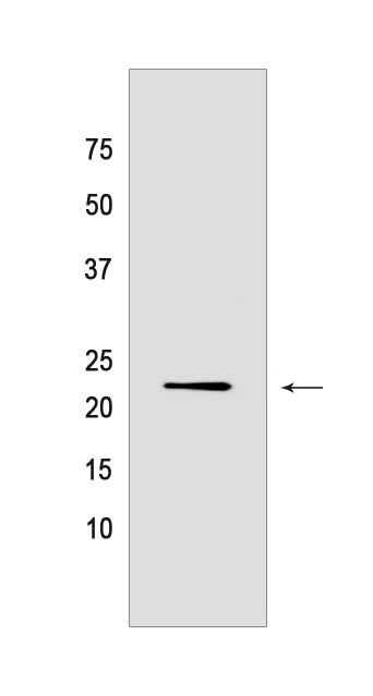
Western blot analysis of extracts from PC12 cells lyastes.using Rho Rabbit mAb [QU7W] at dilution of 1:1000 incubated at 4℃ over night
Product information
Protein names :RHOA,ARH12,ARHA,RHO12,RHOA_HUMAN,Transforming protein RhoA
UniProtID :P61586
MASS(da) :21,768
MW(kDa) :22 kda
Form :Liquid
Purification :Protein A purification
Host :Rabbit
Isotype :IgG
sensitivity :Endogenous
Reactivity :Human,Rat
- ApplicationDilution
- 免疫印迹(WB)1:1000-2000
- 免疫组化(IHC)1:100,
- The optimal dilutions should be determined by the end user
Specificity :Antibody is produced by immunizing animals with a synthetic peptide of Human Rho.
Storage :Antibody store in 10 mM PBS, 0.5mg/ml BSA, 50% glycerol. Shipped at 4°C. Store at-20°C or -80°C. Products are valid for one natural year of receipt.Avoid repeated freeze / thaw cycles.
WB Positive detected :PC12 cells lyastes
Function : Small GTPase which cycles between an active GTP-bound and an inactive GDP-bound state. Mainly associated with cytoskeleton organization, in active state binds to a variety of effector proteins to regulate cellular responses such as cytoskeletal dynamics, cell migration and cell cycle. Regulates a signal transduction pathway linking plasma membrane receptors to the assembly of focal adhesions and actin stress fibers (PubMed:8910519, PubMed:9121475, PubMed:31570889). Involved in a microtubule-dependent signal that is required for the myosin contractile ring formation during cell cycle cytokinesis (PubMed:16236794, PubMed:12900402). Plays an essential role in cleavage furrow formation. Required for the apical junction formation of keratinocyte cell-cell adhesion (PubMed:20974804, PubMed:23940119). Essential for the SPATA13-mediated regulation of cell migration and adhesion assembly and disassembly (PubMed:19934221). The MEMO1-RHOA-DIAPH1 signaling pathway plays an important role in ERBB2-dependent stabilization of microtubules at the cell cortex. It controls the localization of APC and CLASP2 to the cell membrane, via the regulation of GSK3B activity. In turn, membrane-bound APC allows the localization of the MACF1 to the cell membrane, which is required for microtubule capture and stabilization (PubMed:20937854). Regulates KCNA2 potassium channel activity by reducing its location at the cell surface in response to CHRM1 activation,promotes KCNA2 endocytosis (PubMed:9635436, PubMed:19403695). Acts as an allosteric activator of guanine nucleotide exchange factor ECT2 by binding in its activated GTP-bound form to the PH domain of ECT2 which stimulates the release of PH inhibition and promotes the binding of substrate RHOA to the ECT2 catalytic center (PubMed:31888991). May be an activator of PLCE1 (PubMed:16103226). In neurons, involved in the inhibition of the initial spine growth. Upon activation by CaMKII, modulates dendritic spine structural plasticity by relaying CaMKII transient activation to synapse-specific, long-term signaling (By similarity). Acts as a regulator of platelet alpha-granule release during activation and aggregation of platelets (By similarity).., (Microbial infection) Serves as a target for the yopT cysteine peptidase from Yersinia pestis, vector of the plague..
Subcellular locationi :Cell membrane,Lipid-anchor,Cytoplasmic side. Cytoplasm, cytoskeleton. Cleavage furrow. Cytoplasm, cell cortex. Midbody. Cell projection, lamellipodium. Cell projection, dendrite. Nucleus.
IMPORTANT: For western blots, incubate membrane with diluted primary antibody in 1% w/v BSA, 1X TBST at 4°C overnight.
-
About
-
Product
-
Law
-
Literature
-

WeChat scan code follow us