
PDIA6 Rabbit mAb [1Y28]Cat NO.: A46334
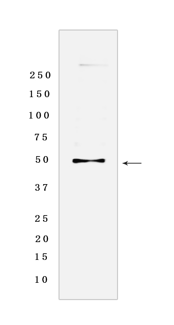
Western blot analysis of extracts from HepG2 cells lyastes.using PDIA6 Rabbit mAb [1Y28] at dilution of 1:1000 incubated at 4℃ over night
Product information
Protein names :PDIA6,ERP5,P5,TXNDC7,PDIA6_HUMAN,Protein disulfide-isomerase A6
UniProtID :Q15084
MASS(da) :48,121
MW(kDa) :48 kDa
Form :Liquid
Purification :Protein A purification
Host :Rabbit
Isotype :IgG
sensitivity :Endogenous
Reactivity :Human,Rat
- ApplicationDilution
- 免疫印迹(WB)1:1000-2000
- 免疫荧光(ICC/IF)1:100,
- The optimal dilutions should be determined by the end user
Specificity :Antibody is produced by immunizing animals with a synthetic peptide of Human PDIA6.
Storage :Antibody store in 10 mM PBS, 0.5mg/ml BSA, 50% glycerol. Shipped at 4°C. Store at-20°C or -80°C. Products are valid for one natural year of receipt.Avoid repeated freeze / thaw cycles.
WB Positive detected :HepG2 cells lyastes
Function : May function as a chaperone that inhibits aggregation of misfolded proteins (PubMed:12204115). Negatively regulates the unfolded protein response (UPR) through binding to UPR sensors such as ERN1, which in turn inactivates ERN1 signaling (PubMed:24508390). May also regulate the UPR via the EIF2AK3 UPR sensor (PubMed:24508390). Plays a role in platelet aggregation and activation by agonists such as convulxin, collagen and thrombin (PubMed:15466936)..
Tissue specificity :Expressed in platelets (at protein level)..
Subcellular locationi :Endoplasmic reticulum lumen. Cell membrane. Melanosome.
IMPORTANT: For western blots, incubate membrane with diluted primary antibody in 1% w/v BSA, 1X TBST at 4°C overnight.
-
About
-
Product
-
Law
-
Literature
-

WeChat scan code follow us