
CLSTN1 Rabbit mAb [L5N4]Cat NO.: A62155
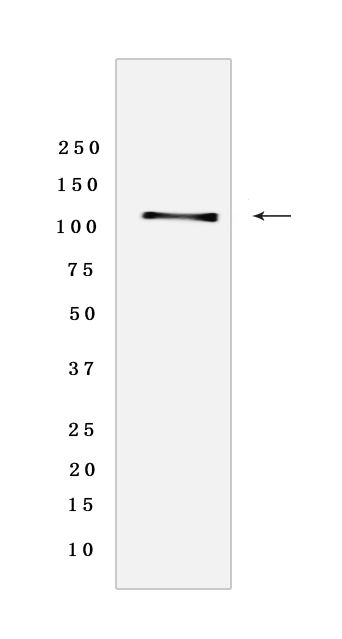
Western blot analysis of extracts from Human fetal brain tissue lyaste.using CLSTN1 Rabbit mAb [L5N4] at dilution of 1:1000 incubated at 4℃ over night
Product information
Protein names :CLSTN1,CS1,KIAA0911,CSTN1_HUMAN,Calsyntenin-1 [Cleaved into: Soluble Alc-alpha ,CTF1-alpha ]
UniProtID :O94985
MASS(da) :109,793
MW(kDa) :110 kDa
Form :Liquid
Purification :Protein A purification
Host :Rabbit
Isotype :IgG
sensitivity :Endogenous
Reactivity :Human,Mouse
- ApplicationDilution
- 免疫印迹(WB)1:1000-2000
- 免疫组化(IHC)1:100,
- 免疫荧光(ICC/IF)1:100,
- The optimal dilutions should be determined by the end user
Specificity :Antibody is produced by immunizing animals with a synthetic peptide of Human CLSTN1.
Storage :Antibody store in 10 mM PBS, 0.5mg/ml BSA, 50% glycerol. Shipped at 4°C. Store at-20°C or -80°C. Products are valid for one natural year of receipt.Avoid repeated freeze / thaw cycles.
WB Positive detected :Human fetal brain tissue lyaste
Function : Induces KLC1 association with vesicles and functions as a cargo in axonal anterograde transport. Complex formation with APBA2 and APP, stabilizes APP metabolism and enhances APBA2-mediated suppression of beta-APP40 secretion, due to the retardation of intracellular APP maturation. In complex with APBA2 and C99, a C-terminal APP fragment, abolishes C99 interaction with PSEN1 and thus APP C99 cleavage by gamma-secretase, most probably through stabilization of the direct interaction between APBA2 and APP. The intracellular fragment AlcICD suppresses APBB1-dependent transactivation stimulated by APP C-terminal intracellular fragment (AICD), most probably by competing with AICD for APBB1-binding. May modulate calcium-mediated postsynaptic signals (By similarity)..
Tissue specificity :Expressed in the brain and, a lower level, in the heart, skeletal muscle, kidney and placenta. Accumulates in dystrophic neurites around the amyloid core of Alzheimer disease senile plaques (at protein level)..
Subcellular locationi :Endoplasmic reticulum membrane,Single-pass type I membrane protein. Golgi apparatus membrane. Cell projection, neuron projection. Cell junction, synapse, postsynaptic cell membrane,Single-pass type I membrane protein. Nucleus.
IMPORTANT: For western blots, incubate membrane with diluted primary antibody in 1% w/v BSA, 1X TBST at 4°C overnight.
-
About
-
Product
-
Law
-
Literature
-

WeChat scan code follow us