
Lgi1/EPT Rabbit mAb [PPX0]Cat NO.: A12491
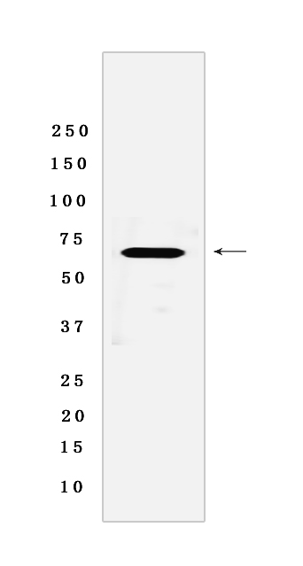
Western blot analysis of extracts from HeLa cells lyastes.using Lgi1/EPT Rabbit mAb [PPX0] at dilution of 1:1000 incubated at 4℃ over night
Product information
Protein names :LGI1,EPT,UNQ775/PRO1569,LGI1_HUMAN,Leucine-rich glioma-inactivated protein 1
UniProtID :O95970
MASS(da) :63,818
MW(kDa) :64 kDa
Form :Liquid
Purification :Protein A purification
Host :Rabbit
Isotype :IgG
sensitivity :Endogenous
Reactivity :Human,Mouse,Rat
- ApplicationDilution
- 免疫印迹(WB)1:1000-2000
- 免疫组化(IHC)1:100,
- The optimal dilutions should be determined by the end user
Specificity :Antibody is produced by immunizing animals with a synthetic peptide of Human Lgi1/EPT.
Storage :Antibody store in 10 mM PBS, 0.5mg/ml BSA, 50% glycerol. Shipped at 4°C. Store at-20°C or -80°C. Products are valid for one natural year of receipt.Avoid repeated freeze / thaw cycles.
WB Positive detected :HeLa cells lyastes
Function : Regulates voltage-gated potassium channels assembled from KCNA1, KCNA4 and KCNAB1. It slows down channel inactivation by precluding channel closure mediated by the KCNAB1 subunit. Ligand for ADAM22 that positively regulates synaptic transmission mediated by AMPA-type glutamate receptors (By similarity). Plays a role in suppressing the production of MMP1/3 through the phosphatidylinositol 3-kinase/ERK pathway. May play a role in the control of neuroblastoma cell survival..
Tissue specificity :Predominantly expressed in neural tissues, especially in brain. Expression is reduced in low-grade brain tumors and significantly reduced or absent in malignant gliomas. Isoform 1 is absent in the cerebellum and is detectable in the occipital cortex and hippocampus,higher amounts are observed in the parietal and frontal cortices, putamen, and, particularly, in the temporal neocortex, where it is 3.5 times more abundant than in the hippocampus (at protein level). Isoform 3 shows the highest expression in the occipital cortex and the lowest in the hippocampus (at protein level)..
Subcellular locationi :Secreted. Cell junction, synapse.
IMPORTANT: For western blots, incubate membrane with diluted primary antibody in 1% w/v BSA, 1X TBST at 4°C overnight.
-
About
-
Product
-
Law
-
Literature
-

WeChat scan code follow us