
IRBIT Rabbit mAb [0W7V]Cat NO.: A34633
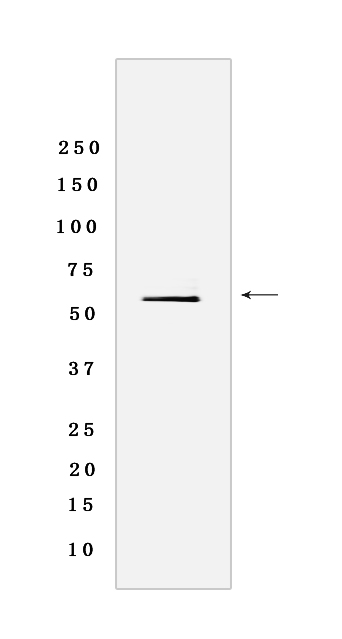
Western blot analysis of extracts from Human thymus tissue lyaste.using IRBIT Rabbit mAb [0W7V] at dilution of 1:1000 incubated at 4℃ over night
Product information
Protein names :AHCYL1,DCAL,IRBIT,XPVKONA,SAHH2_HUMAN,S-adenosylhomocysteine hydrolase-like protein 1 Rs binding protein released with IP)
UniProtID :O43865
MASS(da) :58,951
MW(kDa) :59 kda
Form :Liquid
Purification :Protein A purification
Host :Rabbit
Isotype :IgG
sensitivity :Endogenous
Reactivity :Human
- ApplicationDilution
- 免疫印迹(WB)1:1000-2000
- 免疫荧光(ICC/IF)1:100,
- The optimal dilutions should be determined by the end user
Specificity :Antibody is produced by immunizing animals with a synthetic peptide of Human IRBIT.
Storage :Antibody store in 10 mM PBS, 0.5mg/ml BSA, 50% glycerol. Shipped at 4°C. Store at-20°C or -80°C. Products are valid for one natural year of receipt.Avoid repeated freeze / thaw cycles.
WB Positive detected :Human thymus tissue lyaste
Function : Multifaceted cellular regulator which coordinates several essential cellular functions including regulation of epithelial HCO3(-) and fluid secretion, mRNA processing and DNA replication. Regulates ITPR1 sensitivity to inositol 1,4,5-trisphosphate, competing for the common binding site and acting as endogenous 'pseudoligand' whose inhibitory activity can be modulated by its phosphorylation status. Promotes the formation of contact points between the endoplasmic reticulum (ER) and mitochondria, facilitating transfer of Ca(2+) from the ER to mitochondria (PubMed:27995898). Under normal cellular conditions, functions cooperatively with BCL2L10 to limit ITPR1-mediated Ca(2+) release but, under apoptotic stress conditions, dephosphorylated which promotes dissociation of both AHCYL1 and BCL2L10 from mitochondria-associated endoplasmic reticulum membranes, inhibits BCL2L10 interaction with ITPR1 and leads to increased Ca(2+) transfer to mitochondria which promotes apoptosis (PubMed:27995898). In the pancreatic and salivary ducts, at resting state, attenuates inositol 1,4,5-trisphosphate-induced calcium release by interacting with ITPR1 (PubMed:16793548). When extracellular stimuli induce ITPR1 phosphorylation or inositol 1,4,5-trisphosphate production, dissociates from ITPR1 to interact with CFTR and SLC26A6, mediating their synergistic activation by calcium and cAMP that stimulates the epithelial secretion of electrolytes and fluid (By similarity). Also activates basolateral SLC4A4 isoform 1 to coordinate fluid and HCO3(-) secretion (PubMed:16769890). Inhibits the effect of STK39 on SLC4A4 and CFTR by recruiting PP1 phosphatase which activates SLC4A4, SLC26A6 and CFTR through dephosphorylation (By similarity). Mediates the induction of SLC9A3 surface expression produced by Angiotensin-2 (PubMed:20584908). Depending on the cell type, activates SLC9A3 in response to calcium or reverses SLC9A3R2-dependent calcium inhibition (PubMed:18829453). May modulate the polyadenylation state of specific mRNAs, both by controlling the subcellular location of FIP1L1 and by inhibiting PAPOLA activity, in response to a stimulus that alters its phosphorylation state (PubMed:19224921). Acts as a (dATP)-dependent inhibitor of ribonucleotide reductase large subunit RRM1, controlling the endogenous dNTP pool and ensuring normal cell cycle progression (PubMed:25237103). In vitro does not exhibit any S-adenosyl-L-homocysteine hydrolase activity (By similarity)..
Tissue specificity :Expressed in dendritic cells..
Subcellular locationi :Endoplasmic reticulum. Cytoplasm, cytosol. Apical cell membrane,Peripheral membrane protein. Microsome.
IMPORTANT: For western blots, incubate membrane with diluted primary antibody in 1% w/v BSA, 1X TBST at 4°C overnight.
-
About
-
Product
-
Law
-
Literature
-

WeChat scan code follow us