
IGHD Rabbit mAb [4S59]Cat NO.: A82608
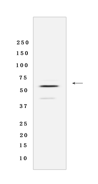
Western blot(SDS-PAGE) analysis of extracts from Human plasma tissue lysate.using IGHD Rabbit mAb [4S59] at dilution of 1:1000 incubated at 4℃ over night.
Product information
Protein names :IGHD,IGHD_HUMAN,Immunoglobulin heavy constant delta
UniProtID :P01880
MASS(da) :42,353
MW(kDa) : 55KDa
Form :Rabbit IgG
Purification :Protein A purification
Host :Rabbit
Isotype :IgG
sensitivity :Endogenous
Reactivity : Human
- ApplicationDilution
- 免疫印迹(WB)1:1000-2000
- 免疫组化(IHC)1:100
- The optimal dilutions should be determined by the end user
Specificity :Antibody is produced by immunizing animals with a synthetic peptide of Human IGHD.
Storage :Antibody store in 10 mM PBS, 0.5mg/ml BSA, 50% glycerol. Shipped at 4°C. Store at-20°C or -80°C. Products are valid for one natural year of receipt.Avoid repeated freeze / thaw cycles.
WB Positive detected : Human plasma tissue lysate
Function : Constant region of immunoglobulin heavy chains. Immunoglobulins, also known as antibodies, are membrane-bound or secreted glycoproteins produced by B lymphocytes. In the recognition phase of humoral immunity, the membrane-bound immunoglobulins serve as receptors which, upon binding of a specific antigen, trigger the clonal expansion and differentiation of B lymphocytes into immunoglobulins-secreting plasma cells. Secreted immunoglobulins mediate the effector phase of humoral immunity, which results in the elimination of bound antigens (PubMed:22158414, PubMed:20176268). The antigen binding site is formed by the variable domain of one heavy chain, together with that of its associated light chain. Thus, each immunoglobulin has two antigen binding sites with remarkable affinity for a particular antigen. The variable domains are assembled by a process called V-(D)-J rearrangement and can then be subjected to somatic hypermutations which, after exposure to antigen and selection, allow affinity maturation for a particular antigen (PubMed:17576170, PubMed:20176268). IgD is the major antigen receptor isotype on the surface of most peripheral B-cells, where it is coexpressed with IgM. The membrane-bound IgD (mIgD) induces the phosphorylation of CD79A and CD79B by the Src family of protein tyrosine kinases. Soluble IgD (sIgD) concentration in serum below those of IgG, IgA, and IgM but much higher than that of IgE. IgM and IgD molecules present on B cells have identical V regions and antigen-binding sites. After the antigen binds to the B-cell receptor, the secreted form sIgD is shut off. IgD is a potent inducer of TNF, IL1B, and IL1RN. IgD also induces release of IL6, IL10, and LIF from peripheral blood mononuclear cells. Monocytes seem to be the main producers of cytokines in vitro in the presence of IgD (PubMed:8774350, PubMed:10702483, PubMed:11282392)..
Subcellular locationi :[Isoform 1]: Secreted.,[Isoform 2]: Cell membrane,Single-pass type I membrane protein.
IMPORTANT: For western blots, incubate membrane with diluted primary antibody in 1% w/v BSA, 1X TBST at 4°C overnight.
-
About
-
Product
-
Law
-
Literature
-

WeChat scan code follow us