
MMP13 Rabbit mAb [1T6T]Cat NO.: A16009
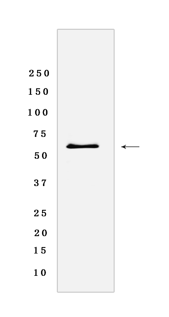
Western blot(SDS-PAGE) analysis of extracts from A-549 cells lysate.using MMP13 Rabbit mAb [1T6T] at dilution of 1:1000 incubated at 4℃ over night.
Product information
Protein names :MMP13,MMP13_HUMAN,Collagenase 3
UniProtID :P45452
MASS(da) :53,820
MW(kDa) :60KDa
Form :Rabbit IgG
Purification :Protein A purification
Host :Rabbit
Isotype :IgG
sensitivity :Endogenous
Reactivity :Human
- ApplicationDilution
- 免疫印迹(WB)1:1000-2000
- 免疫组化(IHC)1:100
- 免疫荧光(ICC/IF)1:100
- The optimal dilutions should be determined by the end user
Specificity :Antibody is produced by immunizing animals with a synthetic peptide of Human MMP13.
Storage :Antibody store in 10 mM PBS, 0.5mg/ml BSA, 50% glycerol. Shipped at 4°C. Store at-20°C or -80°C. Products are valid for one natural year of receipt.Avoid repeated freeze / thaw cycles.
WB Positive detected :A-549 cells lysate
Function : Plays a role in the degradation of extracellular matrix proteins including fibrillar collagen, fibronectin, TNC and ACAN. Cleaves triple helical collagens, including type I, type II and type III collagen, but has the highest activity with soluble type II collagen. Can also degrade collagen type IV, type XIV and type X. May also function by activating or degrading key regulatory proteins, such as TGFB1 and CCN2. Plays a role in wound healing, tissue remodeling, cartilage degradation, bone development, bone mineralization and ossification. Required for normal embryonic bone development and ossification. Plays a role in the healing of bone fractures via endochondral ossification. Plays a role in wound healing, probably by a mechanism that involves proteolytic activation of TGFB1 and degradation of CCN2. Plays a role in keratinocyte migration during wound healing. May play a role in cell migration and in tumor cell invasion..
Tissue specificity :Detected in fetal cartilage and calvaria, in chondrocytes of hypertrophic cartilage in vertebrae and in the dorsal end of ribs undergoing ossification, as well as in osteoblasts and periosteal cells below the inner periosteal region of ossified ribs. Detected in chondrocytes from in joint cartilage that have been treated with TNF and IL1B, but not in untreated chondrocytes. Detected in T lymphocytes. Detected in breast carcinoma tissue..
Subcellular locationi :Secreted, extracellular space, extracellular matrix. Secreted.
IMPORTANT: For western blots, incubate membrane with diluted primary antibody in 1% w/v BSA, 1X TBST at 4°C overnight.
-
About
-
Product
-
Law
-
Literature
-

WeChat scan code follow us