
phospho-p53 (S376) Rabbit mAb [5NH0]Cat NO.: A81882
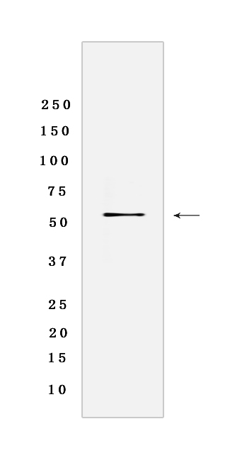
Western blot(SDS-PAGE) analysis of extracts from 293T cells lysate CA-treated.using p53 (S376) Rabbit mAb [5NH0] at dilution of 1:1000 incubated at 4℃ over night.
Product information
Protein names :TP53,P53,P53_HUMAN,Cellular tumor antigen p53
UniProtID :P04637
MASS(da) :43,653
MW(kDa) : 53KDa
Form :Rabbit IgG
Purification :Protein A purification
Host :Rabbit
Isotype :IgG
sensitivity :Endogenous
Reactivity : Human,Rat
- ApplicationDilution
- 免疫印迹(WB)1:1000-2000
- The optimal dilutions should be determined by the end user
Specificity :Antibody is produced by immunizing animals with a synthetic phosphopeptide corresponding to residues around S376 of Human p53.
Storage :Antibody store in 10 mM PBS, 0.5mg/ml BSA, 50% glycerol. Shipped at 4°C. Store at-20°C or -80°C. Products are valid for one natural year of receipt.Avoid repeated freeze / thaw cycles.
WB Positive detected :293T cells lysate CA-treated
Function : Acts as a tumor suppressor in many tumor types,induces growth arrest or apoptosis depending on the physiological circumstances and cell type (PubMed:11025664, PubMed:12524540, PubMed:12810724, PubMed:15186775, PubMed:15340061, PubMed:17317671, PubMed:17349958, PubMed:19556538, PubMed:20673990, PubMed:20959462, PubMed:22726440, PubMed:24051492, PubMed:9840937, PubMed:24652652). Involved in cell cycle regulation as a trans-activator that acts to negatively regulate cell division by controlling a set of genes required for this process (PubMed:11025664, PubMed:12524540, PubMed:12810724, PubMed:15186775, PubMed:15340061, PubMed:17317671, PubMed:17349958, PubMed:19556538, PubMed:20673990, PubMed:20959462, PubMed:22726440, PubMed:24051492, PubMed:9840937, PubMed:24652652). One of the activated genes is an inhibitor of cyclin-dependent kinases. Apoptosis induction seems to be mediated either by stimulation of BAX and FAS antigen expression, or by repression of Bcl-2 expression. Its pro-apoptotic activity is activated via its interaction with PPP1R13B/ASPP1 or TP53BP2/ASPP2 (PubMed:12524540). However, this activity is inhibited when the interaction with PPP1R13B/ASPP1 or TP53BP2/ASPP2 is displaced by PPP1R13L/iASPP (PubMed:12524540). In cooperation with mitochondrial PPIF is involved in activating oxidative stress-induced necrosis,the function is largely independent of transcription. Induces the transcription of long intergenic non-coding RNA p21 (lincRNA-p21) and lincRNA-Mkln1. LincRNA-p21 participates in TP53-dependent transcriptional repression leading to apoptosis and seems to have an effect on cell-cycle regulation. Implicated in Notch signaling cross-over. Prevents CDK7 kinase activity when associated to CAK complex in response to DNA damage, thus stopping cell cycle progression. Isoform 2 enhances the transactivation activity of isoform 1 from some but not all TP53-inducible promoters. Isoform 4 suppresses transactivation activity and impairs growth suppression mediated by isoform 1. Isoform 7 inhibits isoform 1-mediated apoptosis. Regulates the circadian clock by repressing CLOCK-ARNTL/BMAL1-mediated transcriptional activation of PER2 (PubMed:24051492)..
Tissue specificity :Ubiquitous. Isoforms are expressed in a wide range of normal tissues but in a tissue-dependent manner. Isoform 2 is expressed in most normal tissues but is not detected in brain, lung, prostate, muscle, fetal brain, spinal cord and fetal liver. Isoform 3 is expressed in most normal tissues but is not detected in lung, spleen, testis, fetal brain, spinal cord and fetal liver. Isoform 7 is expressed in most normal tissues but is not detected in prostate, uterus, skeletal muscle and breast. Isoform 8 is detected only in colon, bone marrow, testis, fetal brain and intestine. Isoform 9 is expressed in most normal tissues but is not detected in brain, heart, lung, fetal liver, salivary gland, breast or intestine..
Subcellular locationi :Cytoplasm. Nucleus. Nucleus, PML body. Endoplasmic reticulum. Mitochondrion matrix. Cytoplasm, cytoskeleton, microtubule organizing center, centrosome.
IMPORTANT: For western blots, incubate membrane with diluted primary antibody in 1% w/v BSA, 1X TBST at 4°C overnight.
-
About
-
Product
-
Law
-
Literature
-

WeChat scan code follow us