
OXCT1 Rabbit mAb [LOVE]Cat NO.: A44876
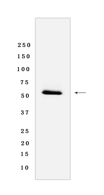
Western blot(SDS-PAGE) analysis of extracts from Rat heart Tissue lyasate.using OXCT1 Rabbit mAb [LOVE] at dilution of 1:1000 incubated at 4℃ over night.
Product information
Protein names :OXCT1,OXCT,SCOT,SCOT1_HUMAN,Succinyl-CoA:3-ketoacid coenzyme A transferase 1, mitochondrial
UniProtID :P55809
MASS(da) :56,158
MW(kDa) :53kDa
Form :Rabbit IgG
Purification :Protein A purification
Host :Rabbit
Isotype :IgG
sensitivity :Endogenous
Reactivity : Human,Mouse,Rat
- ApplicationDilution
- 免疫印迹(WB)1:1000-2000
- 免疫荧光(ICC/IF)1:100
- The optimal dilutions should be determined by the end user
Specificity :Antibody is produced by immunizing animals with a synthetic peptide of Human OXCT1.
Storage :Antibody store in 10 mM PBS, 0.5mg/ml BSA, 50% glycerol. Shipped at 4°C. Store at-20°C or -80°C. Products are valid for one natural year of receipt.Avoid repeated freeze / thaw cycles.
WB Positive detected :Rat heart Tissue lyasate
Function : Key enzyme for ketone body catabolism. Catalyzes the first, rate-limiting step of ketone body utilization in extrahepatic tissues, by transferring coenzyme A (CoA) from a donor thiolester species (succinyl-CoA) to an acceptor carboxylate (acetoacetate), and produces acetoacetyl-CoA. Acetoacetyl-CoA is further metabolized by acetoacetyl-CoA thiolase into two acetyl-CoA molecules which enter the citric acid cycle for energy production (PubMed:10964512). Forms a dimeric enzyme where both of the subunits are able to form enzyme-CoA thiolester intermediates, but only one subunit is competent to transfer the CoA moiety to the acceptor carboxylate (3-oxo acid) and produce a new acyl-CoA. Formation of the enzyme-CoA intermediate proceeds via an unstable anhydride species formed between the carboxylate groups of the enzyme and substrate (By similarity)..
Tissue specificity :Abundant in heart, followed in order by brain, kidney, skeletal muscle, and lung, whereas in liver it is undetectable. Expressed (at protein level) in all tissues (except in liver), most abundant in myocardium, then brain, kidney, adrenal glands, skeletal muscle and lung,also detectable in leukocytes and fibroblasts..
Subcellular locationi :Mitochondrion.
IMPORTANT: For western blots, incubate membrane with diluted primary antibody in 1% w/v BSA, 1X TBST at 4°C overnight.
-
About
-
Product
-
Law
-
Literature
-

WeChat scan code follow us